Injuries to ligaments and tendons
Distortions (sprains) on the arms and legs often occur during sports activities, but also in everyday life. Most events do not lead to serious structural damage, i.e. an injury with permanent consequences. Nevertheless, a torn ligament or tendon can occur. A clinical-manual and imaging examination is usually necessary for a final diagnosis.
Achillodynia
Achillodynia is a painful damage to the Achilles tendon. The disease occurs almost exclusively in athletically active people (especially runners, triathletes, football and tennis players) and is one of the most common sports injuries. It is usually caused by repeated incorrect or excessive strain on the Achilles tendon. Typical for Achillodynia is a load-dependent pain in the area of the rear lower leg and the heel. Non-operative methods (orthopaedic technology and physiotherapy) are usually used for Achillodynia therapy. If the damage is detected and treated in time, its progress can be prevented or at least slowed down.
Torn Achilles tendon
In case of an Achilles tendon rupture (Achilles tendon rupture), the Achilles tendon is completely seperated. This injury is usually caused by overstraining the tendon during sports activities. If the Achilles tendon tears, this is usually associated with a characteristic bang. An Achilles tendon tear typically affects people between the ages of 30 and 50. If the Achilles tendon is torn, this is called a partial Achilles tendon rupture. The Achilles tendon is essential for walking and running. Therefore, if the tendon is torn, a quick and professional surgical or conservative therapy should be carried out.
Therapy
Therapy for a torn Achilles tendon aims to regain the tendon’s resilience and full functionality in the upper ankle joint. Both surgical and non-surgical methods are available, with conservative, non-surgical therapy being used more and more frequently. In the meantime, the affected foot is also no longer immobilised with a plaster cast for several weeks, as is usual for a long time (both after operative and non-operative treatment). This measure has now been replaced by an early, limited exercise therapy, the so-called functional treatment. Special shoes are available for this purpose, which – equipped with a corresponding heel elevation and an inflexible tongue – even allow the affected person to put full weight on the foot after a few days. Since the healing time after an Achilles tendon rupture is at least six weeks, the functional treatment takes just as long.
In order for conservative treatment to be successful, an ultrasound examination (sonography) should be performed by an orthopaedic surgeon experienced in ultrasound immediately after the Achilles tendon rupture (Achilles tendon rupture) to ensure that the ends of the ruptured tendon are in contact with each other when the foot is lowered by about 20 degrees in the so-called pointed foot position. The earlier the non-surgical therapy begins, the greater the chance that it will be successful. It may be necessary to wear a cast for about a week. With the help of special shoes, the patient can put full weight on the foot even after a short time without disturbing the healing process. In the further course of treatment, regular check-ups should be carried out by the orthopaedist, if necessary with an ultrasound examination.
If the Achilles tendon tear (Achilles tendon rupture) is treated surgically, the ends of the tear are sewn together again. After the operation it is necessary to put a plaster cast on the foot for a few days until the skin wound has healed. Afterwards, an early functional treatment, similar to the non-surgical treatment after an Achilles tendon rupture, is possible.
The rupture of the outer ligament is usually caused by indirect trauma in the sense of supination or, more rarely, pronation trauma – popularly known as “buckling”.
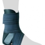
A torn ligament usually does not require surgery. It has been shown that even in cases of severe injury to the external ligament apparatus, a functionally good result can be achieved without surgery. The ankle joint is immobilised for six weeks in a plaster cast, special shoe, splint or orthosis (see illustration). The foot can bear weight as far as the pain allows. The healing process takes about eight to twelve weeks. Sports involving contact, impact or jumping should be avoided for at least 3 months, but in any case until the foot has regained a completely pain-free function.
Torn ligament at the ankle is a partial or complete rupture of one or more ligament structures. The affected joint shows a swelling with pain and bruising. Torn ligaments are usually treated by immobilisation for usually 2-6 weeks or surgically by suturing the ligament or fixing torn pieces of bone.
The ski thumb is the classic ligament injury of the hand, in which the thumb is overstretched outwards, i.e. away from the hand, thereby tearing the ligament on the small finger side at the base joint of the thumb. The task of the ligament is normally to stabilize the so-called bottle handle. If the band were not present, the thumb would bend outwards when grasping a bottle powerfully. This type of ligament tear is most frequently found during skiing: The thumb falls onto the piste or gets caught in the loop of the ski pole. But also the impact of a ball on the floor, during gymnastics, wrestling and self-defense sports can cause a rupture of the ulnar (ulnar side) capsule ligament apparatus at the base of the thumb.
Typical for the “ski thumb” are pain and swelling in the area of the thumb, which is abnormally movable outwards. The first step is to immobilize the thumb with a temporary splint and cool it with ice pads. The joint is unstable when completely torn apart and a full water bottle typically cannot be held in the hand. To exclude fractures or torn bony ligaments, a digital x-ray of the thumb in two planes is obligatory. This can also be carried out as a manual function test in real time under X-ray fluoroscopy (if necessary in lateral comparison) to clarify both bony and ligament injuries or instabilities. In case of a pulled thumb, it is sufficient to immobilize the thumb in a metacarpal thumb orthosis (see figure).
In case of complete instability, however, conservative therapy is not promising and surgical reconstruction is absolutely necessary.


